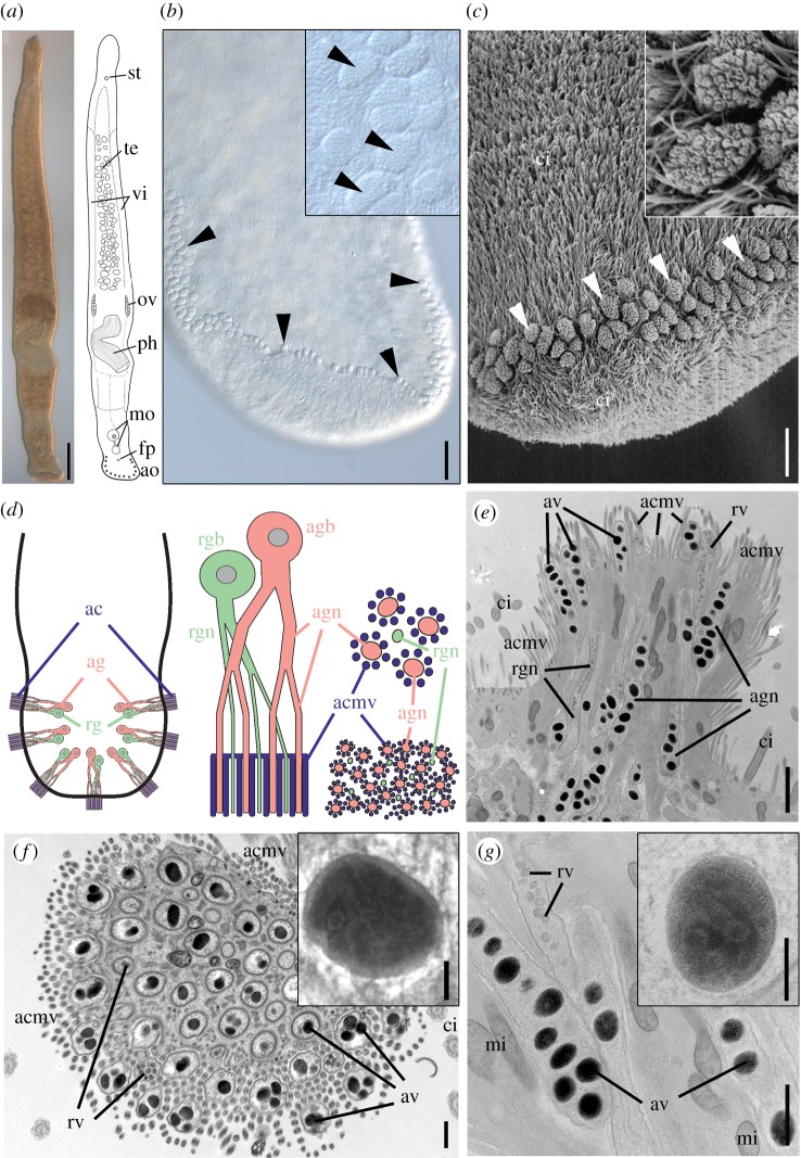
Figure 1.
Morphology of Minona ileanae and the adhesive organs. (a) Differential interference contrast and schematic drawing. (b) Detail of the ventral side of the tail-plate (posterior-most tip slightly folded inside) with adhesive organs (arrowheads); detail of adhesive pads (inset). (c) Scanning electron microscopy of posterior tip of the tail with adhesive organs (arrowheads) and detail (inset). (d) Left: scheme of the organization of adhesive organs in the tail (seven organs shown); middle: scheme of a single adhesive organ with anchor cell (blue), adhesive gland cell (pink), and releasing gland cell (green); bottom right: scheme of a cross-section through the apical region of an adhesive organ (compare with electronic supplementary material, figure S1c,d); top right: detail of four distal ends of adhesive gland cell neck branches (pink) surrounded by microvilli of the anchor cell (blue) and one distal end of a releasing gland cell branch (green). (e–g) Transmission electron micrographs of adhesive organs: (e) longitudinal section from cryo-fixed sample and (f) cross-section through the organ's apical region (chemical fixation). Inset in (f) shows an adhesive vesicle with clear core–periphery zonation of the contents (lead staining). (g) Detail of adhesive and releasing gland cell necks and magnification of one adhesive vesicle with substructures (inset) (uranyl acetate/lead staining of cryo-fixed sample). ac, anchor cell; acmv, anchor cell microvilli; ag, adhesive gland; agn, adhesive gland neck; agb, adhesive gland cell body; ao, adhesive organs; av, adhesive vesicles; ci, cilia; fp, female pore; mi, mitochondria; mo, male organ; ov, ovary; ph, pharynx; rg, releasing gland; rgb, releasing gland body; rgn, releasing gland neck; rv, releasing vesicles; st, statocyst; te, testes; vi, vitellaria. Scale bars (a) 200 µm, (b) 25 µm, (c) 10 µm, (e) 1 µm, (f,g) 500 nm, inset (f,g) 100 nm.

