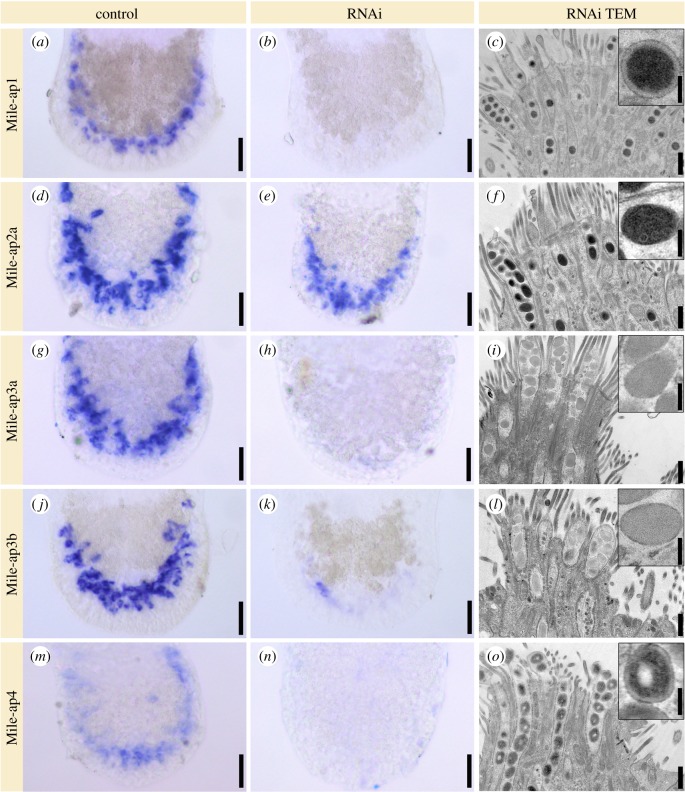
Figure 4.
RNA interference (RNAi) of adhesion candidates. Expression of transcripts in the tails of Minona ileanae. In situ hybridization of control RNAi experiments (first column) and RNAi-treated animals (second column). Third column: transmission electron microscopic images of longitudinal sections of adhesive organs and details of an adhesive vesicle (insets) of RNAi-treated animals. All samples were chemically fixed and sections stained with lead. See text for details. Scale bars 25 µm (all control and RNAi in situ hybridizations), (c) 500 nm and 125 nm (inset) (same for all images of third column).

