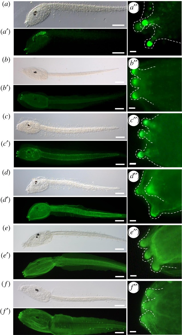
Figure 1.

Enrichment of PNA, PHA-E and GSL II bound carbohydrates in the adhesive organs of Ciona intestinalis larvae. Fluorescent lectin labelling of C. intestinalis larvae with: (a-a″) PNA, (b-b″) PHAL-E, (c-c″) GSL II, (d-d″) DSL, (e-e″) WGA and (f-f ″) ConA. Corresponding bright-field images (a–f), fluorescence (a′–f′) and details of the papillae (a″–f″), highlighted by a dashed line, anterior to the left. Scale bars: (a–f, a′–f ′) 100 μm, (a″–f ″) 10 µm. (Online version in colour.)
