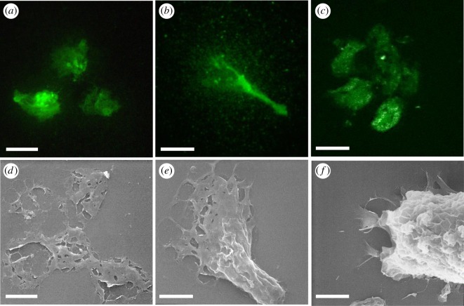
Figure 2.
The presence of PHA-E and GSL II bound carbohydrates in the adhesive plaques of C. intestinalis larvae. (a–c) Confocal images of lectin-fluorescent stainings and (d–f) SEM images of adhesive plaques. (a,b) GSL II and (c) PHAL-E staining. (d,e) SEM pictures of similarly shaped adhesive plaques. (f) Substrate attached larva secreting glue from the adhesive papillae. SEM, scanning electron microscopy. Scale bar: 10 µm.

