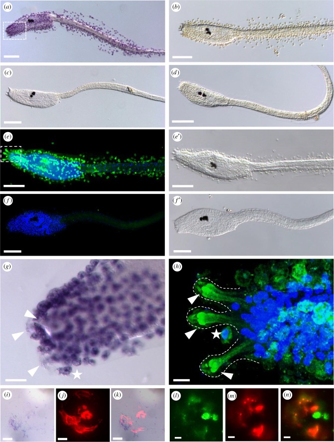
Figure 5.
DOPA presence in test cells, papillae and adhesive plaques of chorion containing larvae. (a–d,g) DOPA-NBT staining and (e-f',h) anti-DOPA-antibody fluorescent staining of Ciona larvae. (a) NBT staining visible in chorionated larvae, is absent upon dechorionation (c) and in controls without substrate (b,d). (e) Double fluorescent labelling with (green) anti-DOPA-antibody and (blue) nuclear DAPI labelling of a chorion containing larva showing the tunic surrounding test cells (e′). (f) Double fluorescent staining of a dechorionated larva having lost both, the DOPA fluorescence and the test cells (f ′). (g,h) Details of DOPA stainings around the adhesive organs in NBT (g) or antibody labelled larvae (h), respectively. The areas of enlargement are marked by rectangles in (a) and (e). Arrowheads mark the papillae, white stars the DOPA-containing cells in papillar vicinity. (i–k) Adhesive plaque staining with NBT (i) and PNA-fluorescence (j), and in the overlay (k). (l–n) Adhesive plaque staining with anti-DOPA-antibody (l) and PNA-fluorescence (m), and in the overlay (n). Dotted lines outline the papillae. Scale bars: (a–h) 100 µm, (g) 20 µm, (h–k) 10 µm. (Online version in colour.)

