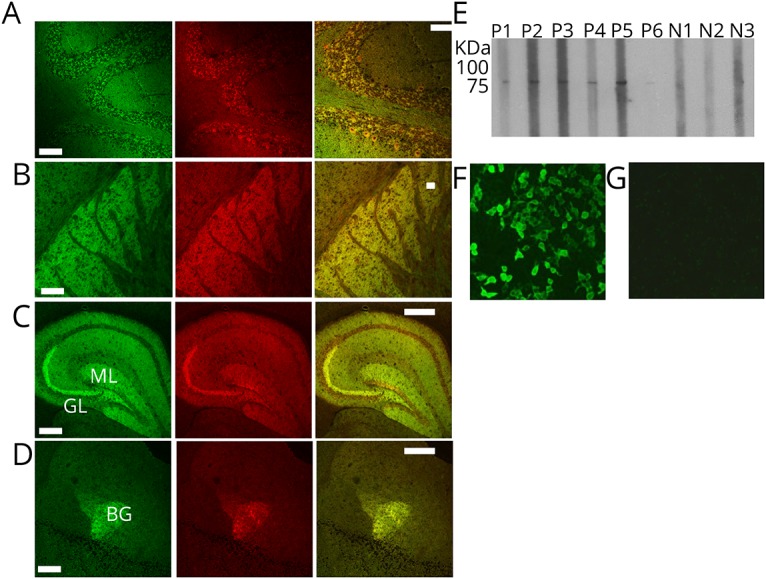
Figure 2. Confocal microscopy and recombinant protein assays (cell-based and Western blot) confirm the antigen is neurochondrin.

(A–D) Patient IgG colocalizes with neurochondrin immunoreactivity in mouse cerebellum, basal ganglia, hippocampus, and the amygdala. Left column: patient IgG is green. Middle column: rabbit antineurochondrin IgG is red. Right column: merged images. (E) In a Western blot utilizing recombinant neurochondrin full-length protein (with 26 kDa GST-tag at N-terminus; total construct molecular weight, 101 kDa), sera from patients 1–6, but not from 3 healthy control subjects, are reactive. (F–G) Patient IgG binding to HEK-293 cells transfected with cDNA encoding neurochondrin (F) and not to nontransfected control cells (G). Scale bar = 100 μm. HEK = human embryonic kidney; cDNA = complementary DNA; IgG = immunoglobulin G.
