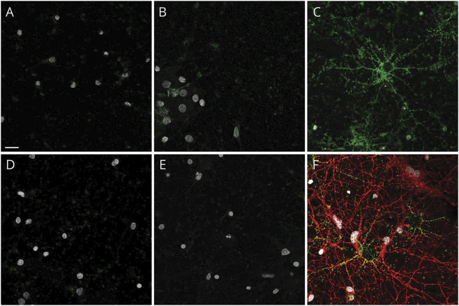
Figure 2. Live cell binding of control and patient immunoglobulin G (IgG) on hippocampal neurons.
(A) Control CSF from normal pressure hydrocephalus patient and (B) healthy control serum do not bind to the surface of living hippocampal neurons. Neither CSF (D) nor serum (E) from patients 1–10 (representative images from patient 5) bind to the neuronal cultures. In contrast, NMDAR-IgG-positive patient CSF binds in a punctate pattern to the extracellular surface of hippocampal neurons (C, green). Cells were poststained for acetylated tubulin to identify axons (F, red). Nuclei stained with DAPI in all panels (white). Scale bar, 20 μm.

