
Fig. 7.
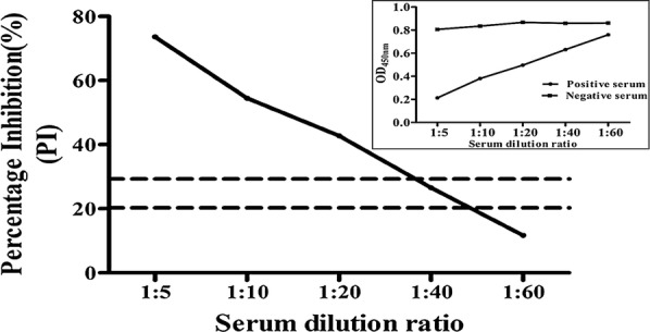
Sensitivity analysis of the blocking ELISA. A 96-well plate was coated with the PEDV N protein (100 ng/well) and then blocked. Serially diluted positive and negative sera were added to the wells and incubated at 37 °C for 2 h. Then, biNb2 was added to the wells, and HRP-conjugated streptavidin was added to the wells. Values are the mean PI (%) of three well replicated. The small figure in the upper right corner shows the OD450nm value of the positive and negative sera. The dotted lines represent the upper and lower cutoff values