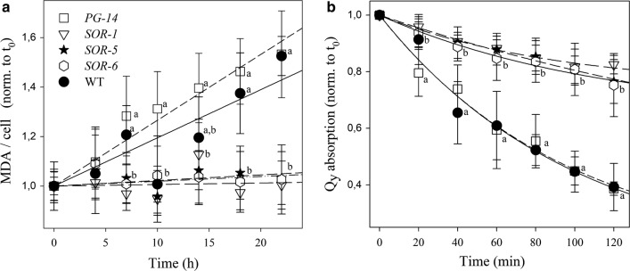
Fig. 4.
Photooxidation of C. vulgaris WT, PG-14 and SOR mutant genotypes under photooxidative stress. a Cell suspensions were treated with 1400 µmol photons m−2 s−1 at 20 °C, and kinetics of malondialdehyde (MDA) formation were followed. MDA is an index of membrane lipid peroxidation, and was quantified by HPLC as thiobarbituric reactive substances. (B) Cell suspension of WT and mutant strains were treated with strong white light (14,000 µmol photons m−2 s−1, 20 °C) and the amount of Chl was evaluated by measuring the absorption area in the region 600–750 nm. See “Materials and methods” for details. Symbols and error bars show mean ± SD, n = 4. Values marked with the same letters are not significantly different from each other within the same time point (ANOVA, p < 0.05)

