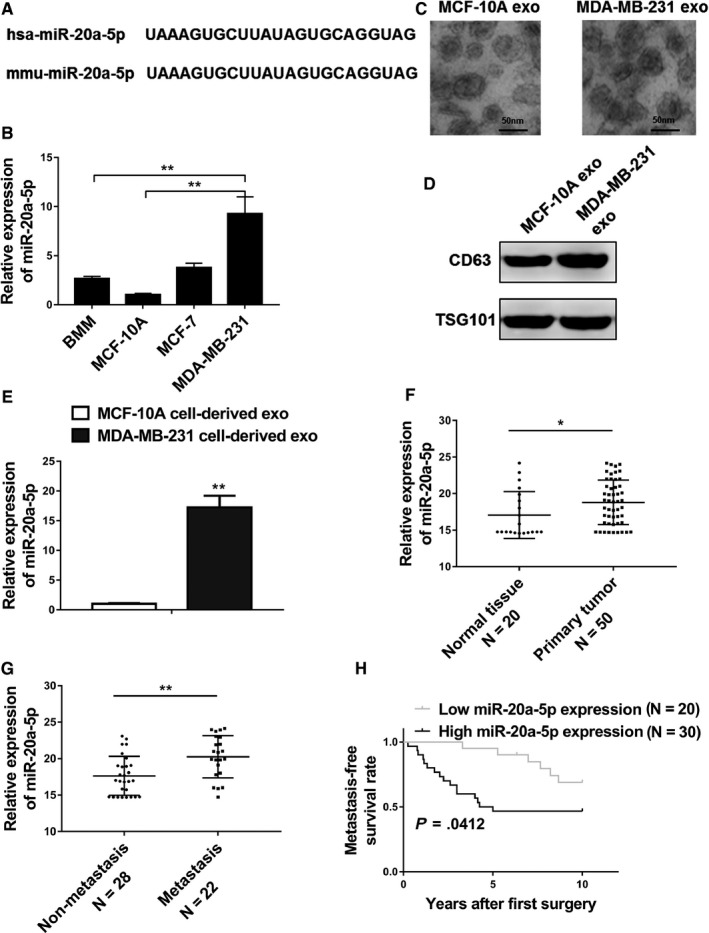
Figure 1.

MiR‐20a‐5p was highly expressed in breast tumor tissues and the exosomes of MDA‐MB‐231 cells. A, A comparison of nucleotides of the mature miR‐20‐5p in humans and mice. B, qRT‐PCR analysis revealed miR‐20‐5p expression in primary murine bone marrow macrophages (BMMs), MCF‐10A cells, MCF‐7 cells, and MDA‐MB‐231 cells. C, Representative electron micrographs of exosomes isolated from MCF‐10A cell conditioned and MDA‐MB‐231 cell conditioned medium revealing the typical morphology and size (50‐150 nm), bar = 50 nm. D, Western blot analysis showing abundant CD63 and TSG101 in exosomes derived from the medium of MCF‐10A (MCF‐10A exo) and MDA‐MB‐231 (MDA‐MB‐231 exo) cells. E, qRT‐PCR analysis revealing miR‐20‐5p expression in MCF‐10A cell‐derived and MDA‐MB‐231 cell‐derived exosomes. F, Expression levels of miR‐20‐5p in 20 normal tissues and 50 breast cancer tissues. G, Relative expression levels of miR‐20‐5p in groups of breast cancer tissues classified based on the occurrence of bone metastasis (metastatic or nonmetastatic). H, Kaplan‐Meier's analysis of the correlation between miR‐20‐5p expression and the metastasis‐free survival of breast cancer patients. The data represent the mean ± SD from three independent experiments. *P < .05; **P < .01
