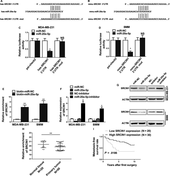
Figure 5.

MiR‐20a‐5p promoted osteoclastogenesis by targeting SRCIN1. A and B, Predicted binding sites of miR‐20a‐5p in the wild type 3′UTR of SRCIN1 (SRCIN1 3′UTR) and mutations in the 3′UTR of SRCIN1 (SRCIN1 3′UTR‐mut) in humans and mice. C and D, The luciferase activities in MDA‐MB‐231 and BMMs cotransfected with indicated miR‐20a‐5p mimics or its negative control mimics (miR‐NC) and constructed luciferase reporter vectors (SRCIN1 3′UTR, SRCIN1 3′UTR‐mut, psi‐check2) were detected as the relative ratio of hRluc luciferase activity to hluc + luciferase activity. E, Detection of SRCIN1 mRNAs in biotinylated miRNA/target mRNA complex by real‐time RT‐PCR. The relative level of SRCIN1 mRNA in the complex pulled down by using biotinylated miR‐20a‐5p was compared to that of the complex pulled down by using the biotinylated control random RNA. F and G, The relative expression levels of SRCIN1 in MDA‐MB‐231 and BMMs cells transfected with indicated microRNA mimics or microRNA inhibitors detected by qRT‐PCR and Western blot. H, Expression levels of SRCIN1 in 20 normal tissues and 50 breast cancer tissues. I, Kaplan‐Meier's analysis of the correlation between SRCIN1 expression and the metastasis‐free survival of breast cancer patients. The data represent the mean ± SD from three independent experiments. *P < .05; **P < .01
