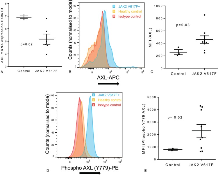
Figure 1.
AXL is upregulated and activated in MPN; CD34+ cells were enriched from control and JAK2 V617F patients using CliniMACS (Miltenyi Biotec). mRNA expression levels for AXL (A) in CD34+ cells from control and JAK2 V617F patients were measured by qRT-PCR and expressed as delta ct values (mean ± SEM, n = 3 normals, n = 6 for JAK2 V617F). CD34+ cells isolated from normal and JAK2 V617F patients were stained with anti AXL (B,C) and anti Y779 phospho-AXL (D,E) antibody and expression levels analyzed on a Novocyte flow cytometer (ACEA Biosciences) using FloJo software. Representative FACS plots are shown (B and D) and amalgamated data (C and E) displayed as median fluorescent intensity ± SEM (n = 3 normals, n = 9 for JAK2 V617F). The results of a t test are shown.

