
Figure 4.
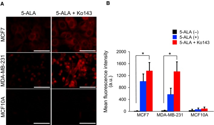
Enhancement of PpIX‐FI with Ko143 in breast cancer cells. (A), Representative confocal fluorescence images of PpIX after incubation with 5‐ALA (5 mmol/L for 2 h) with or without Ko143 (1 μmol/L). Bars = 50 μm. (B), Mean FI of PpIX for 2 h of incubation. All error bars represent SD (n = 3). *P < .05