Figure 1.
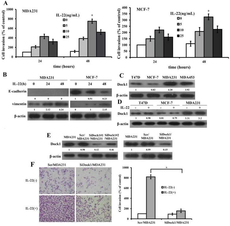
IL-22 promoted invasion and EMT of breast cancer cells. Dock1 was required for IL-22-mediated MDA-MB-231 cells invasion and EMT. A. The IL-22-triggered invasion of breast cancer cells was analyzed. Left, quantitative analysis of MDA-MB-231 cells under diverse concentrations of IL-22 (0-25 ng/mL) with time gradience. Right, quantitative analysis of MCF-7 cells under diverse concentrations of IL-22 (0-25 ng/mL) with time gradience. *P< 0.05. B. Level of E-cadherin and Vimentin in MDA-MB-231 and MCF-7 cells with and without 10 ng/mL IL-22 was examined by western blot at different time points. C. Expression of Dock1 protein in cultured breast cancer cells. D. Levels of Dock1 protein in breast cancer cells were assessed by western blot after 48 hours with and without stimulation of 10 ng/mL IL-22. E. Western blot analysis of Dock1 expression in indicated cells. F. The IL-22-induced invasion of MDA-MB-231 cells was evaluated. Left, photographs of invading cells (magnification, 200×). Right, quantification of invading cells. *P < 0.05. For B, C, D, E, data of western blot were representative of triply repeated experiments. Used β-actin as loading control. Quantifications of relative protein level were shown below the blots.

