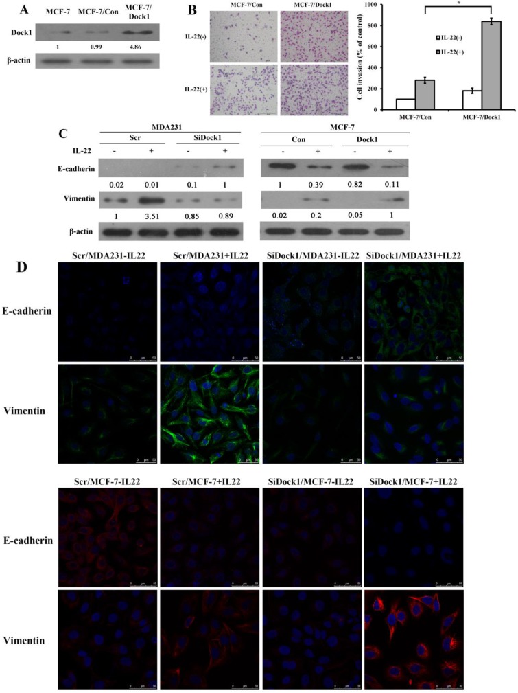
Figure 2.
Dock1 was required for IL-22-mediated MCF-7 cell invasion, metastasis, and EMT. A. Western blot analysis of Dock1 expression in indicated cells. B. The IL-22-induced invasion of MCF-7 cells was assessed. Left, photographs of invading cells (magnification, 200×). Right, quantification of invading cells. *P< 0.05. The results shown were representative of triply repeated experiments. C. Western blot of E-cadherin level and Vimentin level in indicated cells after 48 h with or without 10 ng/mL IL-22. D. Fluorescence microscopy of stained E-cadherin and Vimentin presented in indicated cells after 48h with IL-22. Nuclear DNA was stained with DAPI (blue). Scale bar: 50 μm. For A and C, western blot results were representative from triply repeated experiments. Used β-actin as loading control. Quantifications of relative protein level are shown below the blots.

