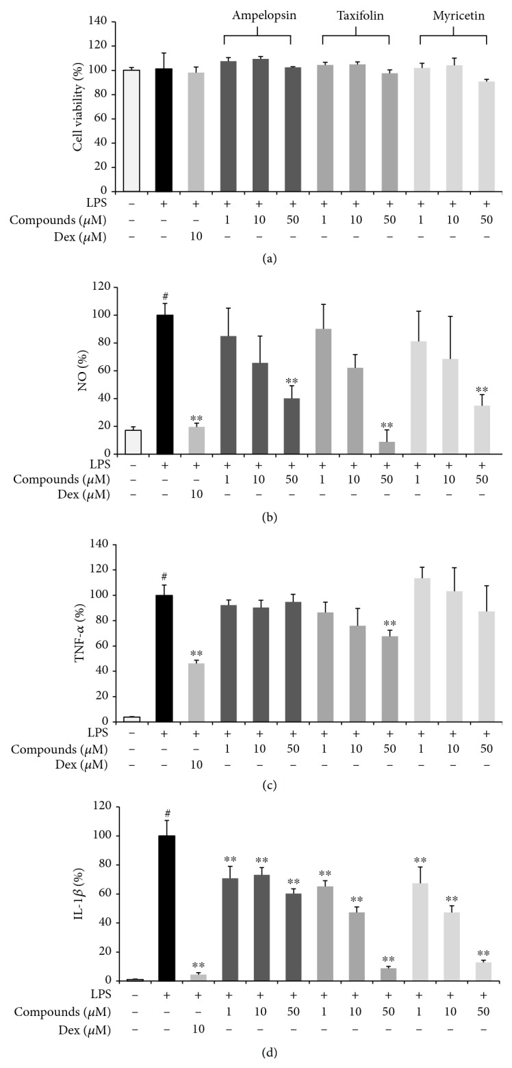
Figure 9.

Effects of three compounds, ampelopsin, taxifolin, and myricetin, on (a) cell viability and the secretion of (b) NO and (c–e) inflammatory cytokines. Cells were seeded with (a, b) 5.0 × 10⁴ cells/well on a 96-well culture plate or (c–e) 2.5 × 10⁵ cells/well on a 24-well culture plate and preincubated for 18 h. Then, cells were pretreated with each compound for 1 h and stimulated with LPS for another 24 h. At least three independent tests were repeated to ensure reproducibility of the experimental results. (a) Cell viability was examined using a cell-counting kit. (b) NO secretion into the culture media was determined using the Griess assay. (c–e) Secretion of inflammatory cytokines was measured by ELISA. As a control, cells were incubated with vehicle alone. Data represent the mean ± SEM of duplicate determinations from three independent experiments. #P < 0.001 (vs. control) and ∗∗P < 0.001 (vs. LPS) values were considered statistically significant.
