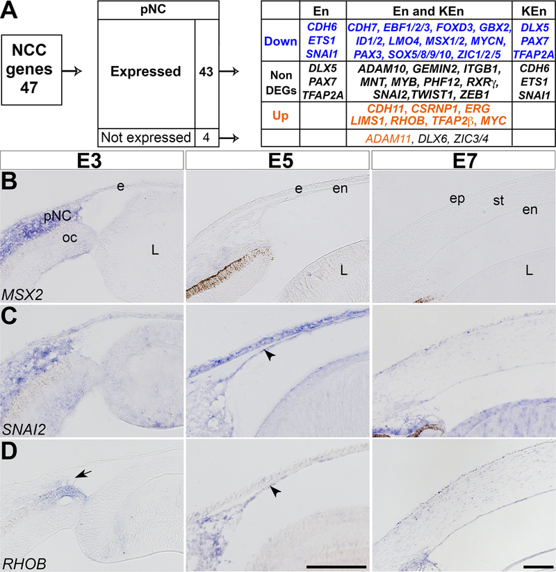
Figure 2. Changes in the molecular identity of NCC during their localization in the periocular region and differentiation into corneal cells.

(A) Analysis based on 47 candidate NCC genes indicates that 43 genes were expressed and 4 were not expressed in the periocular region. Of the 43 expressed genes, 20 were downregulated, 10 were constitutively expressed, and 7 were upregulated during corneal development. (B-D) Section in situ hybridization of E3, E5, and E7 anterior eyes indicating that: (B) MSX2 is strongly expressed in the periocular region but undetectable in the corneal endothelium and stroma; (C) SNAI2 is maintained at all three time points; and (D) RHOB is minimal in the periocular region, but it is strongly expressed in the corneal endothelium and stroma. Arrow indicates periocular region and arrowheads indicate corneal endothelium. Abbreviations: pNC, periocular neural crest; ec, ectoderm; oc, optic cup; L, lens; ep, epithelium; en, endothelium; st, stroma. Scale bars: 100 μm.
