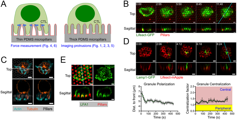
Figure 1. CTLs form protrusions on stimulatory micropillars.
(A) Schematic diagram of thin micropillars used for force measurements (left) and thicker micropillars used for imaging protrusions (right). (B-E) OT1 CTLs were imaged by confocal microscopy on micropillars bearing H2-Kb-OVA and ICAM1. z-projection images (top views) are shown above with sagittal views below. Dotted lines (cyan in B, D, and E, white in C) denote the slicing plane used for the sagittal images. (B) Time-lapse montage of a representative CTL expressing Lifeact-GFP, with micropillars shown in red. (C) Fixed image of a representative CTL stained with phalloidin (to visualize F-actin) and anti-tubulin antibodies. Micropillars are shown in gray in the left images. (D) Above, time-lapse montage of a representative CTL expressing Lifeact-mApple and Lamp1-GFP. Below left, mean distance between the lytic granule cloud and the cell front, graphed against time. Below right, centralization factor analysis of Lamp1-GFP. In both graphs, time 0 denotes initial contact with the pillars and error bars indicate standard error of the mean (SEM). N = 10. (E) Fixed image of a representative CTL stained with anti-LFA1 antibodies, with micropillars shown in red. All scale bars = 2 μm. In B and D, time in M:SS is indicated in the upper left corner of each top view image.

