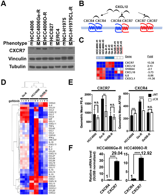
Figure 3. CXCR7 and CXCL12 are overexpressed in gefitinib-resistant HCC4006Ge-R cells with a mesenchymal phenotype.
A. Immunoblot showing CXCR7 expression with vinculin and β-tubulin as loading controls. B. Schematic diagram showing CXCL12-mediated activation of CXCR4 or CXCR7. C. A heat map for the overexpressed chemokine and cytokine genes (FDR<0.05, FC>3) in mesenchymal HCC4006Ge-R cells and epithelial HCC4006GeSB-R cells compared to parental HCC4006 cells. D. A heatmap showing relative concentration of cytokine and chemokines in HCC4006, HCC4006 Ge-R, or HCC4006GeSB-R cells treated with DMSO (−) or 500nmol/L gefitinib (+). E. Cell surface expression of CXCR7 or CXCR4 in cells expressing shRNAs against Non-Target (ΔNT) or CXCR7 (ΔCX). Average of three independent assays, (**p≤0.01, ***p≤0.001, and p<0.0001, N.S., not significant). F. The estimated expression ratio between CXCR4 and CXCR7 mRNAs in HCC4006Ge-R cells or HCC4006O-R cells calculated using GUSB as an internal control. Average of three independent assays, (****p≤0.0001).

