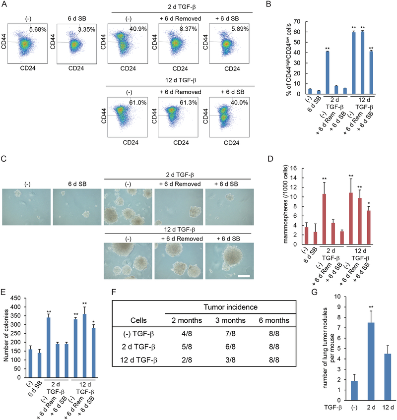
Figure 5. Prolonged TGF-β treatment stabilizes a stem cell state and induces latency in breast cancer cells.
(A and B) HMLER cells were treated with TGF-β for 2 or 12 days (d), followed by its removal, as indicated, and then cultured with (SB) or without (Rem) SB431542 for 6 days. The expression of cell surface markers CD44 and CD24 was analyzed by flow cytometry (A). The graph (B) shows the percentages of CD24lowCD44high stem cells in the entire cell population. Data are mean ± S.E. from N=3 independent experiments. **P< 0.01 by a Dunnet’s test. (C and D) HMLER cells were treated as in (A) and were assessed for mammosphere formation. The mammospheres were observed by phase-contrast microscopy (C) and counted (D). Scale bar, 100 μm. Data are mean ± S.E. from N=3 independent experiments. *P< 0.05 and **P< 0.01 by a Dunnet’s test. (E) HMLER cells were treated as in (A) and assessed for colony formation in soft agar. Data are mean ± S.E. from N=3 independent experiments. *P< 0.05 and **P< 0.01 by a Dunnet’s test. (F) HMLER cells were treated with TGF-β for 0, 2 or 12 days then suspended in PBS with 50% Matrigel, and 10,000 cells were injected orthotopically into a mammary fat pad of NSG mice. The number of mice with a palpable tumor is indicated in the table. (G) HMLER cells were treated with TGF-β for 0, 2 or 12 days then suspended in PBS, and 500,000 cells were injected into tail vein of NSG mice. Six weeks after injection, the mice were sacrificed, and lungs were harvested. Cancer dissemination into the lungs was measured by counting the tumor nodules in the lungs. Data are mean ± S.E. from N=3 independent experiments with N=7 mice. **P< 0.01 by a Dunnet’s test.

