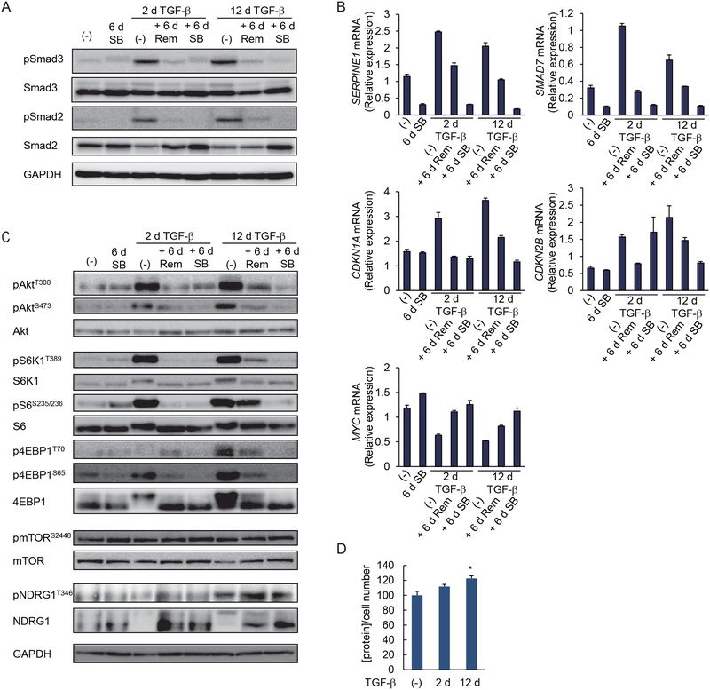
Figure 6. Prolonged TGF-β treatment enhances and stabilizes AKT-mTOR signaling.
(A) HMLER cells were treated with TGF-β for 2 or 12 days (d), followed by its removal, as indicated, and were cultured in the media without added TGF-β (Rem) or with SB431542 (SB) for 6 days. C-terminal phosphorylation of Smad3 and Smad2 was analyzed by immunoblotting. Blots are representative of N=3 independent experiments. (B) HMLER cells were treated as in (A), and the expression of TGF-β target genes encoding PAI1, Smad7, p21Cip1, p15Ink4B or c-Myc was assessed by qRT-PCR and normalized to rPL19 mRNA. Data are mean ± S.E. from N=3 independent experiments. (C) HMLER cells were treated as in (A) and phosphorylation of AKT, S6K1, S6, 4EBP1, mTOR and NDRG1 was analyzed by immunoblotting. Blots are representative of N=3 independent experiments. (D) HMLER cells were treated with TGF-β for 0, 2 or 12 days. The cells were trypsinized and counted. The total protein content normalized for cell number is shown relative to untreated cells. Data are mean ± S.E. from N=3 independent experiments. *P< 0.05 by a Dunnet’s test.

