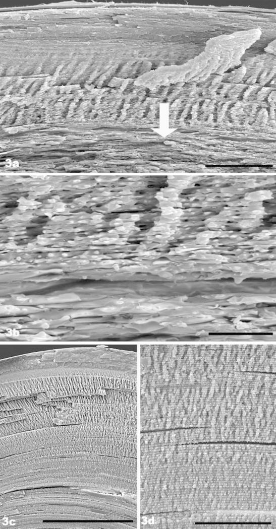
FIGURE 3.
(a) SEM of 7.5-month-old filensin knockout. The lens surface at the bow region is located at the top. The most superficial region of the “hand” zone is evident. However, the loss of structure caused by the absence of beaded filaments begins a short way into the hand zone (arrow), greatly reducing its domain (compare with Fig. 4b of wild type). (b) Higher magnification SEM view of the region, where the impact of BF absence first becomes evident in the 7.5-month-old knockout (a region comparable to the area overlaid by the arrow in Fig. 3a). (c) SEM overview of 7.5-month-old wild-type lens, showing the normal, much larger extent of the hand domain. Black bar, 200 μm. In contrast to (a), the long-range, highly ordered stacking of fiber cells is evident well into the deep cortex. (c) Higher magnification view of the deep cortex of the 7.5-month-old wild-type lens, showing the gradual transition from the hand domain to deeper cortex. This contrasts with the abrupt loss of fiber cell shape and regular packing evident in the knockout (a, b). Scale bar: (a), 50 μm; (b) 10 μm; (c) 50 μm.

