Figure 1.
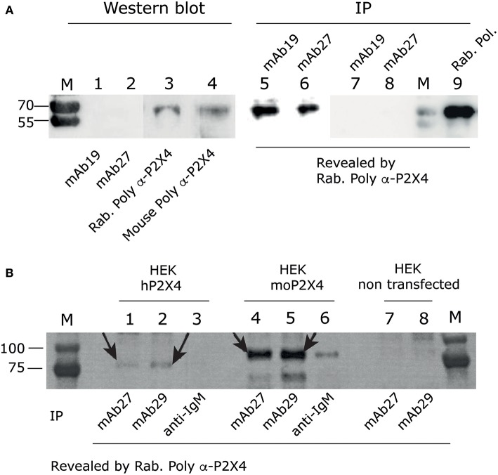
Anti-hP2X4 recognizes hP2X4 in immunoprecipitation experiments, but not in western blot. (A) Left panel. Western blot using mAb19 (1), mAb27 (2), rabbit anti-rat-P2X4 polyclonal antibodies from Alomone (3) or our mouse anti-hP2X4 polyclonal antibodies (4), performed on total cell lysate of HEK293 cells transfected with hP2X4. Blots were revealed using anti-mouse and anti-rabbit HRP Abs. Right panel. hP2X4 was immuno-precipitated from the same lysate of HEK293 cells expressing human P2X4, using mAb19 (5) or mAb27 (6), and revealed by western blotting with rabbit anti-P2X4 polyclonal antibodies. Negative controls are shown in lanes 7 and 8: immunoprecipitation of non transfected HEK293 cell lysates with mAb19 (lane 7) and mAb27 (lane 8). Immunoprecipitation of P2X4 from cell lysate of HEK293 cells transfected with hP2X4 using rabbit anti-rat P2X4 polyclonal antibodies is shown in lane 9. (B) Immunoprecipitation assays using mAb27 (IgG2b; lanes 1, 4, 7) or mAb29 (IgM; lanes 2, 5, 8) or anti-IgM (lanes 3 and 6) from HEK cells overexpressing hP2X4-mcherry (lanes 1–3), mouse P2X4-mcherry (lanes 4–6), or non transfected (lanes 7, 8). P2X4 was revealed by western blotting with rabbit anti-P2X4 polyclonal antibodies. Arrows indicate hP2X4 or moP2X4 bands.

