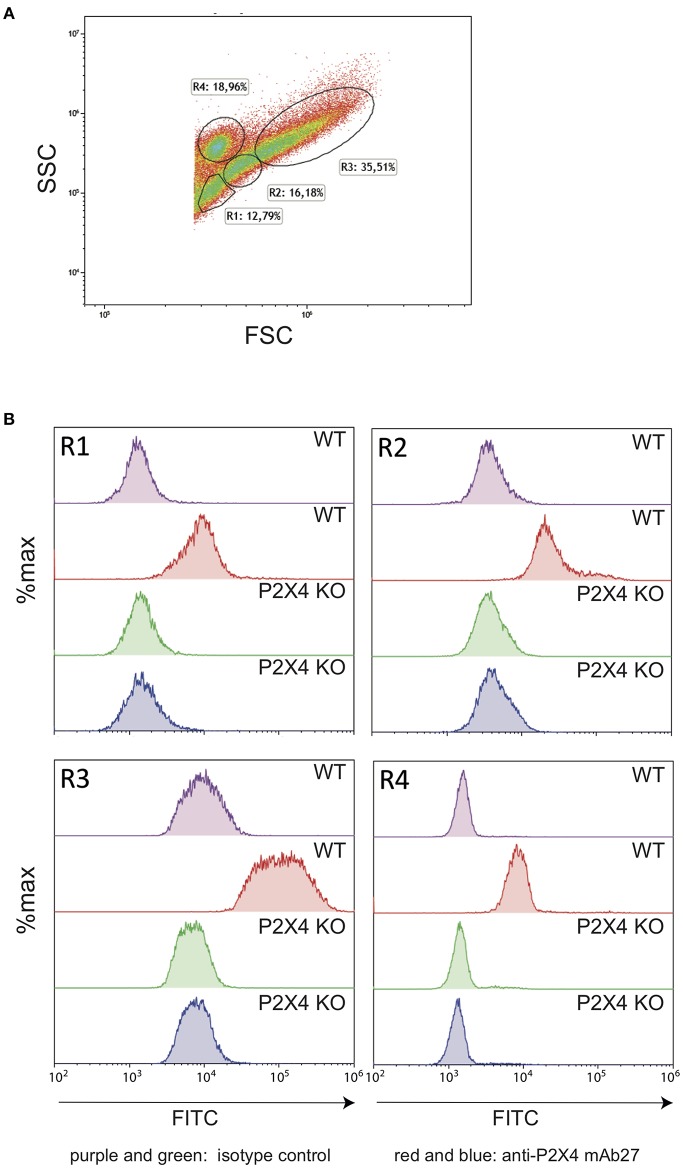
Figure 2.
Anti-hP2X4 mAb27 recognizes the mouse P2X4 specifically. (A) FSC/SSC representation of mouse peritoneal cells (C57BL/6 strain). Gates were defined as in Hermida et al. (32). Erythrocytes were gated out. During the preparation of cells, erythrocytes were not lysed to avoid ATP release, which would trigger P2X4. Each cell suspension corresponds to a pool from three different mice of each genetic background. (B) Comparison of wild type (WT) and P2X4 KO peritoneal cells from gates R1–R4 labeled with isotype control (purple, WT and green, P2X4 KO) or with mAb27 FITC (red, WT and blue, P2X4 KO).

