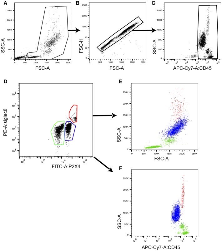
Figure 7.
Eosinophils from human PBL express highest levels of P2X4. CD45+ peripheral blood leukocytes defined by the gating strategy presented in (A–C) were stained with fluorochrome-coupled anti-P2X4 mAb27 and with anti-Siglec8 mAb, and analyzed by flow cytometry. A representative dot plot shows P2X4 vs. SIGLEC8 expression (D) in CD45+ leukocytes. Three gates outlined in red (gate 1), blue (gate 2), and green (gate 3) define P2X4highSIGLEC8high, P2X4medSIGLEC8low, and P2X4lowSIGLEC8low subsets, respectively. (E,F) Represent forward (FSC) (respectively, CD45 expression) vs. side scatter (SSC) for cells from each gate, showing that P2X4highSIGLEC8high are large, highly granular cells.

