Figure 8.
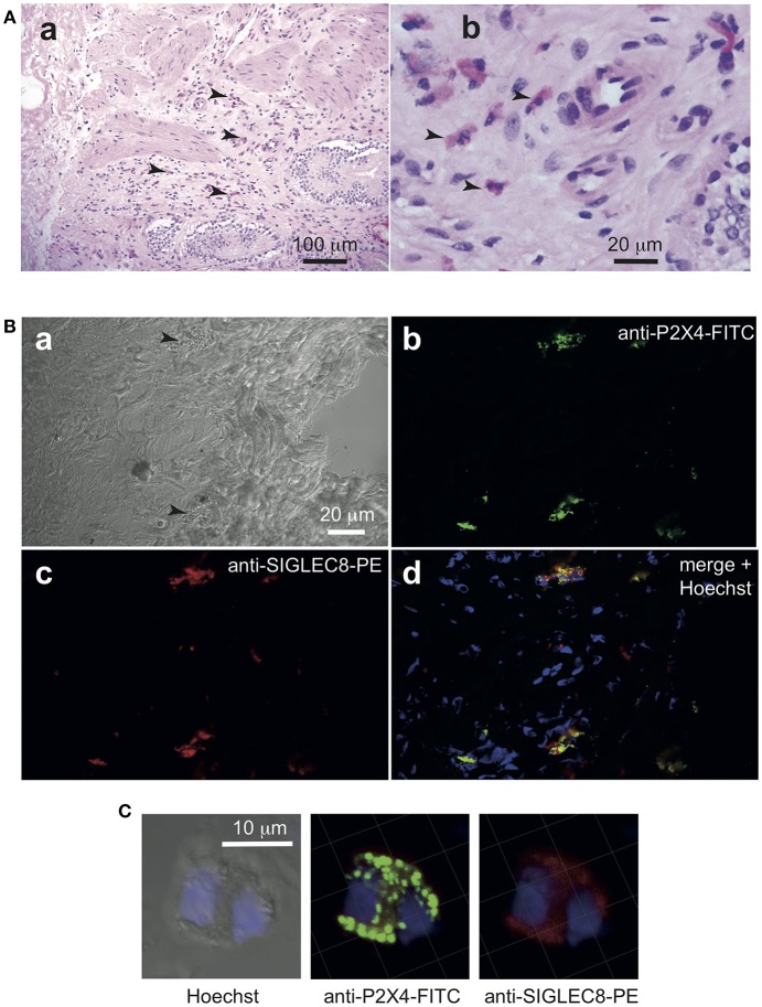
Anti-hP2X4 staining identifies eosinophils in gall bladder sections. Cryosections (5 μm) of freshly isolated gall bladder sample from a patient with cholecystitis diagnosis were stained by hematoxylin-eosin (A). Eosinophils are indicated by arrows. In (B), two granulocytes are indicated by black arrows in the bright field image (a). Sections were stained with anti-hP2X4-FITC mAb27 (b), anti Siglec-8-PE (c). Merged images with Hoechst staining is shown in (d). Images acquired by confocal microscopy indicate that Siglec8-positive eosinophils express high levels of P2X4. (C) Illustrates the respective intracellular distributions of Siglec-8 and P2X4. Isotypic control is shown in Figure S5.

