
Fig. 3. Tetrode trials and behavioral study in rats.
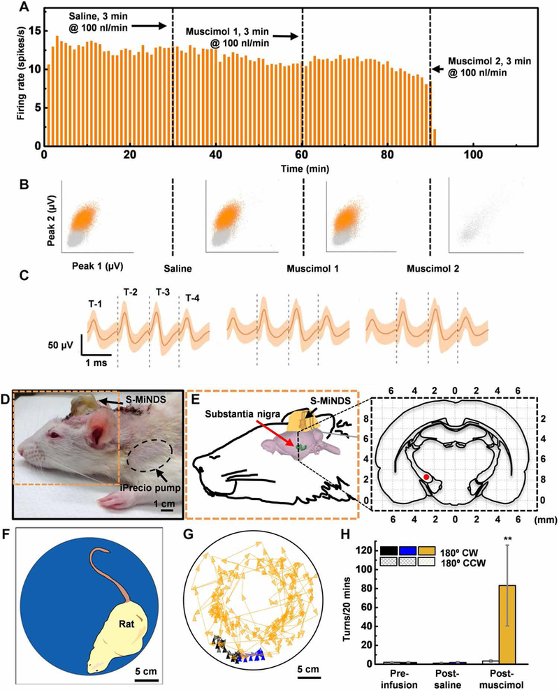
(A) Firing rate histograms for 1-min bins of the recorded unit. The vertical black dashed lines indicate the start of saline infusion (first line at 30 min), the muscimol infusion (second line at 60 min), and the second muscimol infusion (third line at 90 min), respectively (n = 1). (B) Representations of sorted (orange) and unsorted (gray) action potentials based on peak values. Peaks 1 and 2 are the maximal value of waveforms measured by T-1 and T-2, respectively, during each period (preinfusion baseline, 22,928 waveforms; saline infusion, 20,940 waveforms; muscimol 1 infusion, 19,073 waveforms; and muscimol 2 infusion, 132 waveforms; n = 1 unit). (C) Averaged action potentials of a well-isolated unit (single unit) during each period (saline infusion, 20,940 wave-forms; muscimol 1 infusion, 19,073 waveforms; and muscimol 2 infusion, 132 waveforms; n = 1 unit). Orange shading represents a band around the mean with a width of three SDs. (D and E) Picture and schematic illustration (orange dashed outline) of a rat with an implanted MiNDS targeting substantia nigra. Expanded view of black dashed line in (E) shows coronal cross section of the brain 5-mm posterior to the bregma. The red dot identifies the position of the MiNDS tip within substantia nigra. (F) Schematic of a rat inside an opaque chamber during the behavioral study. (G) Color-tracking map of a rat during the preinfusion, post-saline, and post-muscimol infusions periods. (H) Mean number of 180° clockwise (CW) and counterclockwise (CCW) turns during preinfusion, post-saline, and post-muscimol infusion periods (n = 3 rats, 2 trials per rat; error bars represent SE; **P < 0.0021). Statistical analysis was done using one-way ANOVA followed by Tukey post hoc test.