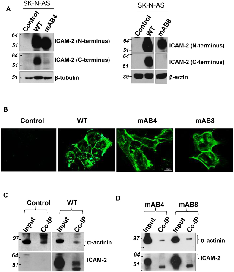
Figure 3. ICAM-2 WT and variants localized to cell membranes.
(A) Immunoblots confirmed that tranfected SK-N-AS cells expressed readily detectable levels of ICAM-2 WT, mAB4 or mAB8. Data in both membranes were generated in the same experiment, but immunoblotted individually. (B) Immunofluorescence staining demonstrated that ICAM-2 WT, mAB4 and mAB8 localized to cell membranes. Scale bar represents 10 μm for all panels. Details of procedures used are in the Methods. (C, D) ICAM-2 WT, but not mAB4 and mAB8, co-precipitated with α-actinin. Immunoprecipitations (IP) were performed using whole cell lysates and a mouse monoclonal antibody to α-actinin (MAB1682, Millipore-Fisher Scientific). The presence of all three types of ICAM-2 proteins in the “input” whole cell lysates (wcl) was confirmed prior to IP.

