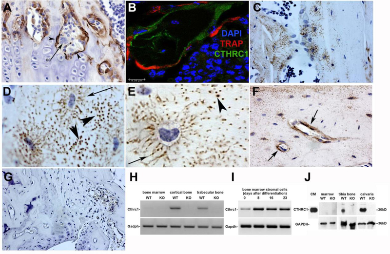
Figure 1. CTHRC1 is expressed in bone by osteoblasts and osteocytes, not osteoclasts.
(A) Monoclonal anti-CTHRC1 antibody reveals CTHRC1 in osteoblasts (arrowheads) and osteocytes of trabecular and cortical bone in wildtype mice, whereas multinucleated osteoclasts (arrows) do not express CTHRC1. (B) Confocal imaging of TRAP (red) and CTHRC1 (green) immunohistochemistry of a bone trabeculum shows no overlap, demonstrating that CTHRC1 is not expressed in cells expressing the osteoclast marker TRAP (nuclear stain with DAPI). (C) Note the distinct localization of CTHRC1 in canaliculi of some osteocytes. (D, E) CTHRC1 is found in the thin cytoplasmic processes of osteocytes (arrow). Immunoreactive CTHRC1 appears to be present in the wider canaliculi (arrows) indicating secretion of CTHRC1 into the interstitial fluid. (F) Prominent accumulation of CTHRC1 is frequently seen around venules (arrows). (G) CTHRC1 is not detectable in trabecular bone of Cthrc1 knockout mice demonstrating antibody specificity. (H) Cthrc1 expression was analyzed by RT-PCR using mRNA isolated from bone fractions of metaphysis and proximal epiphysis (trabecular bone), diaphysis (cortical bone), as well as bone marrow, which does not express Cthrc1. (I) Cthrc1 mRNA expression was analyzed in primary bone marrow-derived mesenchymal stromal cells during osteogenic differentiation. (J) Western blot analysis of CTHRC1 from femur and marrow lysates obtained from wildtype and Cthrc1 null mice shows CTHRC1 in bone but not the marrow fraction.

