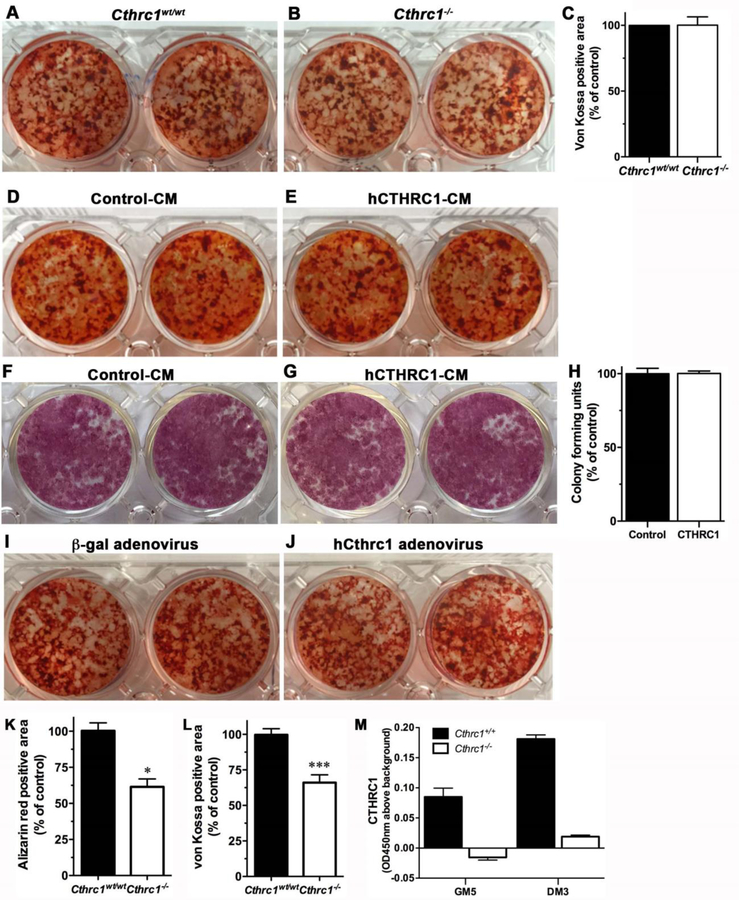
Figure 3. CTHRC1 does not affect osteogenic differentiation of bone marrow derived stromal cells or bone formation in vitro.
Alizarin red staining shows similar mineral content in bone marrow-derived stromal cells isolated from (A) wildtype or (B) Cthrc1 null mice after induction of osteogenic differentiation. (C) Bone marrow stromal cell cultures from wildtype and Cthrc1−/− mice form similar amounts of bone in vitro as determined by von Kossa staining. (D, E) Alizarin red staining (D, E) and alkaline phosphatase activity staining (F, G) show that differentiation of bone marrow-derived stromal cells isolated from Cthrc1−/− mice in the presence of control conditioned-medium (Control-CM) or hCTHRC1 (hCTHRC1-CM) is similar. von Kossa staining showed similar results (not shown). (H) Bone marrow stromal cell proliferation was unaffected by the presence or absence of CTHRC1 with similar numbers of colonies formed. (I, J) Following transduction with control adenovirus (b-gal) or hCTHRC1 adenovirus bone marrow-derived stromal cells isolated from Cthrc1 null mice revealed similar osteogenic differentiation potential. (K, L) Calvarial osteoblast cell cultures from Cthrc1 null mice showed reduced bone formation as determined by decreased Alizarin red and von Kossa staining. (M) CTHRC1 was secreted into the medium by wildtype osteoblast cultures as determined by ELISA (GM5- day 5 in growth medium, DM3- day 3 in differentiation medium). Differentiation was performed over a 20-day period for all experiments. *p<0.05, ***p<0.005.

