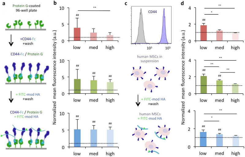
Figure 2. Modification of HA influences CD44 binding to HA macromers in soluble form.
(a) Protein G-coated plates were treated with a soluble CD44-Fc chimera for directional presentation of CD44. The plate surface was then exposed to FITC-modified HA macromers and the resulting fluorescent signal was quantified to assess (b) the influence of the extent and type of modification on CD44-HA interactions (red: NorHA1, green: NorHA2, blue: MeHA). (c) Human MSCs that express CD44 (shown through flow cytometry) were exposed to FITC-modified HA macromers and analyzed using flow cytometry to determine (d) the influence of the extent and type of modification on interactions between cell-surface CD44 and modified HA (red: NorHA1, green: NorHA2, blue: MeHA). n=8 for surface measurements and n=3 for flow measurements per group, dotted lines represent PEG controls, * P < 0.05; ** P < 0.01; ## P< 0.01 relative to PEG control.

