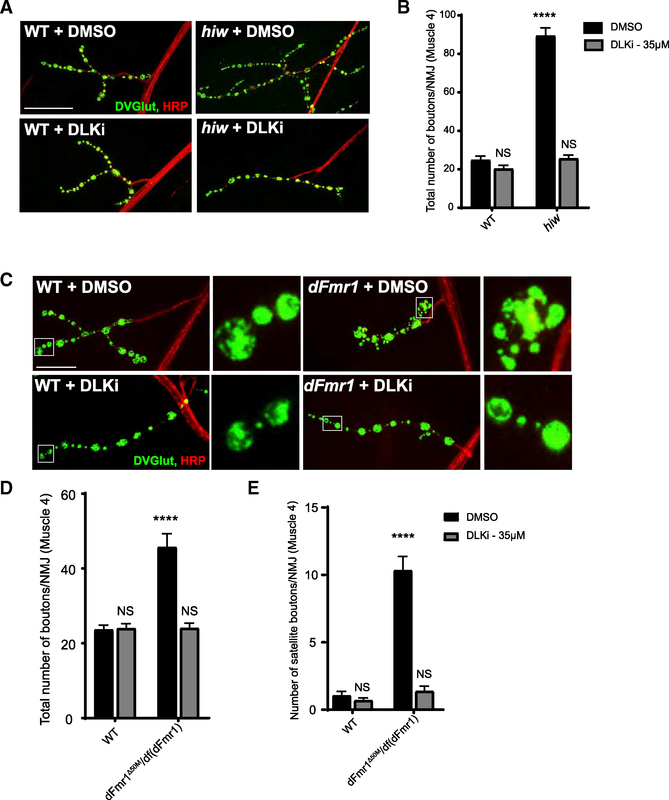
Figure 6. An Inhibitor Against the Mammalian Ortholog of Wnd, DLK, Suppresses NMJ Morphology Defects in dFmr1 Mutant Larvae.
(A) Representative images of the NMJ synaptic terminal at muscle 4 in third-instar wild-type and hiw mutant larvae raised on food treated with either DMSO (vehicle control) or DLK inhibitor (DLKi). NMJs were stained for the presynaptic bouton marker DVGLUT (green) and nerve membrane marker HRP (red). Scale bar: 50 μm.
(B) Quantification of the mean (±SEM) number of boutons per muscle 4 NMJ in each genotype in each treatment condition. Feeding the DLK inhibitor to larvae is sufficient to suppress Wnd-dependent hiw synaptic overgrowth.
(C) Representative images of the NMJ synaptic terminal at muscle 4 in third-instar wild-type and dFmr1 mutant larvae raised on food treated with either DMSO (vehicle control) or DLK inhibitor. Representative bouton images are shown in the inset panels to the right of the respective genotype. Scale bar: 25 μm.
(D) Quantification of the mean (±SEM) number of total boutons per muscle 4 NMJ in each genotype in each treatment condition.
(E) Quantification of the mean (±SEM) number of satellite boutons per muscle 4 NMJ in each genotype. Feeding the DLK inhibitor to dFmr1 larvae is sufficient to suppress increases in total and satellite bouton number.
*p = 0.05; **p = 0.01; ***p = 0.001; ****p = 0.0001; NS, not significant; p > 0.05. p values shown on figure represent statistical comparison to wild type treated with DMSO.
Detailed statistical tests and additional comparisons available in Table S1.

