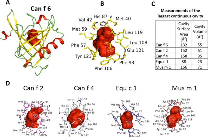
Fig 6. A comparison of the ligand binding cavities in the different lipocalins.

Cavity residues and volume maps (red) were calculated using a rolling probe of 1.4Å and displayed as Connolly surfaces (25, 26, 27). (A) The overall ligand core is centrally located within the beta barrel and closed at the top by the L1 and L5 loops. (B) Residues from multiple strands of the β-barrel contribute to the ligand cavity core with hydrophobic amino acids and aromatic side chains in the interior of the ligand binding surface. (C) The ligand cavity volumes are fairly similar except for Equ c 1. (D) The ligand cavities of Can f 2 (red), Can f 4 (grey), Equ c 1 (purple), and Mus m 1 (brown) have a variable shape. Can f 6 and Equ c 1 have similar ligand cavity shape, with the slight differences between them likely reflects slightly differently shaped ligands.
