Figure 2.
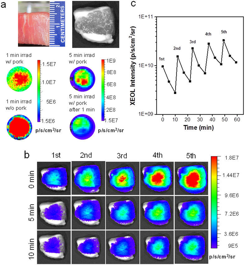
XEOL imaging from beneath deep tissues using LGO:Cr nanoparticles. a) Stimulation of XEOL from under 1.5-cm thick pork. Pork slice was put on top of the particles during X-ray irradiation but removed during imaging. Left: afterglow images taken 1 min after X-ray irradiation. Right: afterglow images taken 5 min after X-ray irradiation. b) XEOL can be detected from under 1.5-cm thick pork. The images were acquired immediately after as well as 5 min and 10 min after X-ray exposure. The pork slice was remained on top of the particles throughout the experiment. c) XEOL intensity changes, based on BLI imaging results from b).

