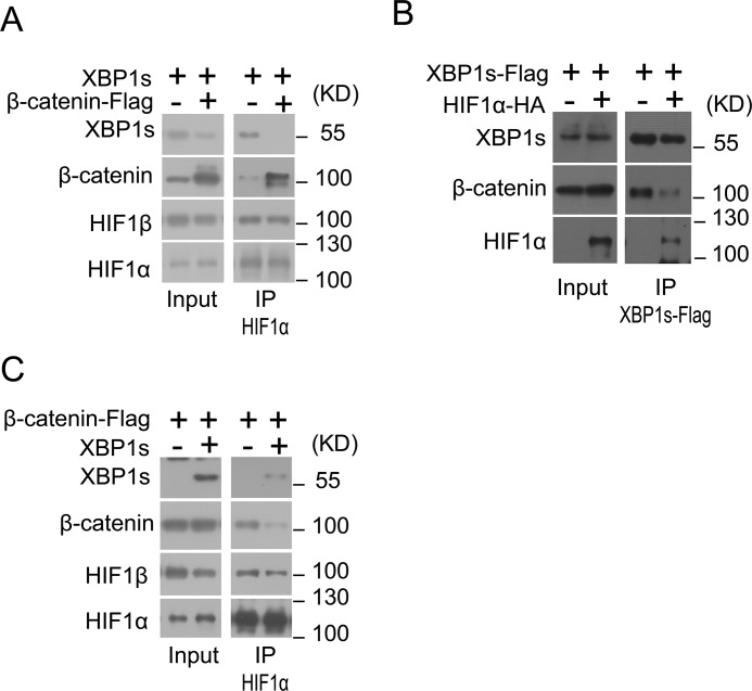
Figure 5.
β-Catenin interacts with XBP1s and HIF1α. A, HEK293 cells were co-transfected with XBP1s overexpression plasmid together with empty vector (−) or β-catenin–FLAG plasmid for 24 h and were then cultured under hypoxia (1% O2) for 12 h. Cell lysates were immunoprecipitated (IP) with anti-HIF1α antibody and analyzed by immunoblotting using the indicated antibodies. B, HEK293 cells were co-transfected with XBP1s-FLAG plasmid along with empty vector (−) or HIF1α–HA plasmid for 24 h and cultured under normoxia conditions. Cell lysates were immunoprecipitated with anti-FLAG antibody and analyzed by immunoblotting using the indicated antibodies. C, HEK293 cells were co-transfected with β-catenin–FLAG plasmid along with empty vector (−) or XBP1s plasmid for 24 h and were subsequently cultured under hypoxia (1% O2) for 12 h. Cell lysates were immunoprecipitated with anti-HIF1α antibody and analyzed by immunoblotting using the indicated antibodies. All results represent three independent experiments.

