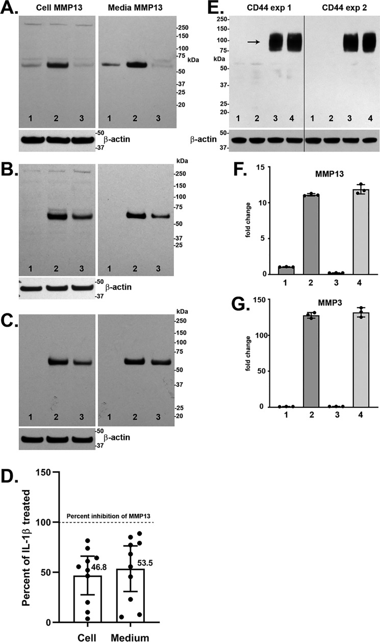
Figure 2.
A–C, Western blot analyses of cell lysate and medium aliquots from three representative experiments with human OA chondrocytes. Chondrocytes were transduced with Ad-Tet-mycHAS2 and subsequently treated without (lane 1) or with (lanes 2 and 3) 1.0 ng/ml IL1β in the absence (lanes 1 and 2) or presence (lane 3) of 100 ng/ml Dox. Blots were probed for detection of MMP13 protein (∼54 kDa), and the cell lysate blots were reprobed for β-actin. The bar graph in D depicts the mean ± 95% confidence interval (error bars) of 10 independent experiments showing the changes in cell-associated and medium accumulation of MMP13 protein due to HAS2-OE (percent inhibition) relative to values with IL1β treatment (without Dox) set to 100% (dotted line). Mean values are shown beside each bar. E, Western blot analyses for CD44 from two representative experiments on bovine chondrocytes transduced with Ad-Tet-human-CD44 and treated with 100 ng/ml Dox (lanes 3 and 4), without IL1β (lanes 1 and 3), or with 1.0 ng/ml IL1β (lanes 2 and 4). Shown is RT-PCR analysis for MMP13 (F) and MMP3 (G) from no Dox (bars 1 and 2) or CD44-OE chondrocytes (bars 3 and 4) treated without (bars 1 and 3) or with 1.0 ng/ml IL1β (bars 2 and 4). Shown is a representative experiment (n = 3) of three independent experiments with similar results. No change in MMP13 or MMP3 mRNA was observed with CD44-OE.

