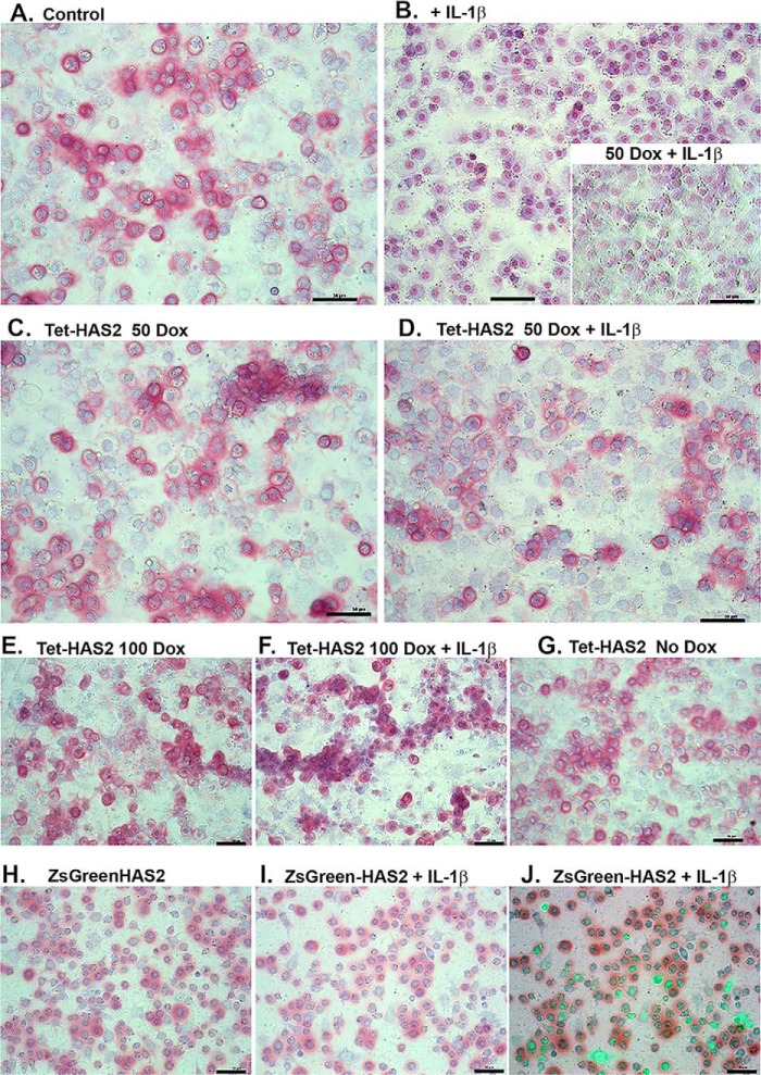
Figure 4.
To determine the effects of IL1β and HAS2-OE at a functional level, control bovine chondrocytes (A and B), chondrocytes transduced with Ad-Tet-murine-mycHAS2 (C–G), or Ad-ZsGreen-human-HAS2 (H–J) were then treated without or with IL1β (1 ng/ml) and varying concentrations of Dox (as labeled) for 1 week. B (inset), nontransduced chondrocytes co-treated with IL1β and 50 ng/ml Dox. The cultures were then fixed and stained with DMMB, a dye that provides pink staining of accumulated proteoglycan (as well as a bluish counterstaining of nuclei). Images shown are representative fields of four independent experiments. J, overlay image of green fluorescence (due to Ad-ZsGreen-mycHAS2 transduction) and bright field. For this image, the green fluorescence intensity has been digitally enhanced to better illustrate successfully transduced cells. Bars in each image, 50 μm.

