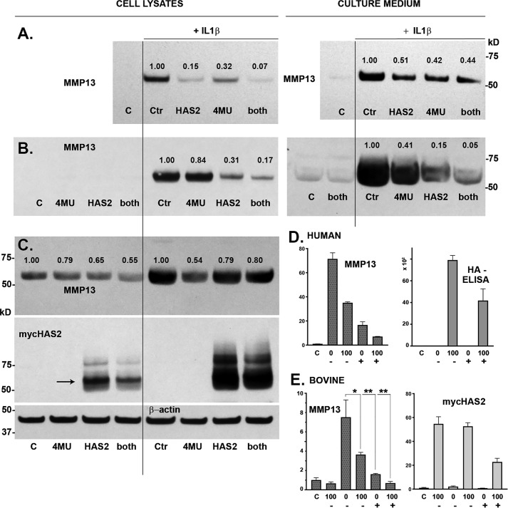
Figure 5.
To determine the independent and combined effects of HAS2-OE and 4MU, chondrocytes were transduced with Ad-Tet-mycHAS2 and subsequently incubated with or without IL1β (1 ng/ml) and co-treated in the absence (control (C)) or presence of 100 ng/ml Dox (labeled HAS2 and both), and/or 0.5 mm 4MU (labeled 4MU and both). The effects of these treatments on MMP13 protein are shown in A–C, representing Western blot analysis of three independent preparations of human OA chondrocytes. A and B, MMP13 present in cell lysates and medium (as labeled); C includes visualization of the mycHAS2 protein and a representative blot of β-actin (β-actin for studies in A and B not shown). Numbers shown above MMP13 bands indicate the relative band intensity (normalized to β-actin) as compared with the intensity of the IL1β-induced MMP13 band set to 1.0. D, a representative example of human OA chondrocytes analyzed for MMP13 mRNA as well as HA content present in the medium (units = ng/ml × 102). Chondrocytes were treated with IL1β (bars 2–5) and co-treated with DOC (0 or 100 as labeled) and 4MU (depicted as − or +). MMP13 data depict the relative -fold change in mRNA relative to control (C) set to 1.0. E, changes in relative expression of MMP13 and mycHAS2 mRNA in bovine chondrocytes. Chondrocytes were treated with IL1β (bars 2–6) and co-treated with DOC (0 or 100 as labeled) and 4MU (depicted as − or +). MMP13 data depict the relative -fold change in mRNA relative to control (C) set to 1.0. E summarizes results from three independent bovine cultures for each condition. An unpaired t test was used for statistical analysis: *, p < 0.05; **, p < 0.01. Error bars, S.D.

