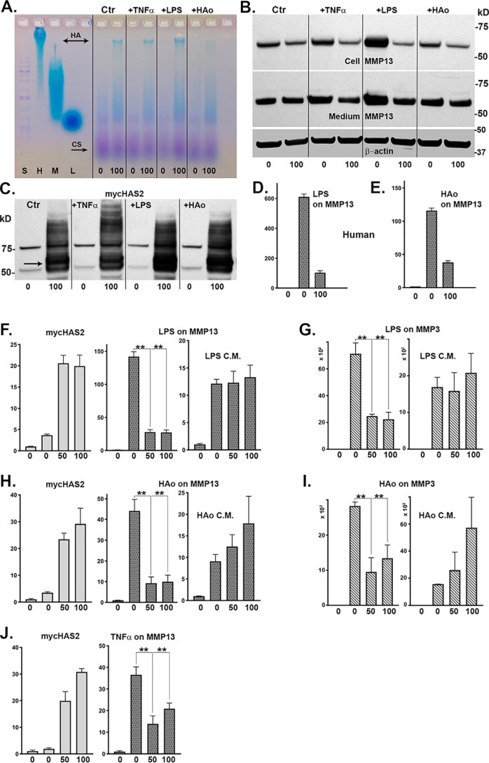
Figure 7.
Bovine or human chondrocytes were activated by other known pro-inflammatory/DAMP agents: TNFα, LPS, or HAo. Ad-Tet-mycHAS2-transduced human chondrocytes, treated with 100 ng/ml Dox as labeled, exhibited enhanced synthesis of high-molecular-mass HA (A) under control (Ctr) and agent-treated conditions. B, representative Western blot analysis of lysates from these same treated cultures probed for MMP13 (blots reprobed for β-actin) and for expression of mycHAS2 protein (C). Next, human chondrocytes were transduced with Ad-Tet-mycHAS2 and then incubated with or without Dox (as labeled 0 and 100); without (bar 1) or with (bars 2 and 3) 10 ng/ml LPS (D) or 250 μg/ml HAo (E). D and E, relative -fold change in MMP13 mRNA, assayed in duplicate, with untreated control values set to 1.0. Bovine chondrocytes transduced with Ad-Tet-mycHAS2 (F–J) were incubated with 0, 50, or 100 ng/ml Dox (as labeled) and without (bar 1) or with (bars 2–4) co-treatment with LPS (F and G), HAo (H and I), or 10 ng/ml TNFα (J). After cultures were exposed to LPS and HAo, the 24-h conditioned medium (LPS C.M. or HAo C.M.) was collected and added to fresh bovine monolayer cultures for an additional 24-h incubation, similar to conditions described in the legend to Fig. 6. Changes in mycHAS2, MMP13, or MMP3 mRNA were quantified. F–J, results from three independent bovine chondrocyte cultures for each condition (each assayed in duplicate). The -fold changes in mRNA are relative to control values set to 1.0. An unpaired t test was used for statistical analysis: **, p < 0.01. Error bars, S.D.

