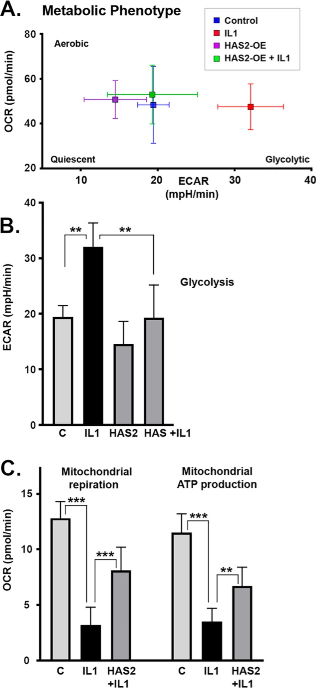
Figure 8.

To determine whether the inhibitory effects of HAS2-OE on MMPs was due to changes in intracellular metabolism, bovine chondrocytes were transduced with Ad-Tet-mycHAS2 and then plated into 24-well (A) or 96-well (B and C) Seahorse XF or XFe cell culture microplates. The confluent monolayers were then incubated with 0, 50, or 200 ng/ml Dox, without or with 1 ng/ml IL1β as labeled. The cells were then mated with a sensor cartridge and analyzed in a Seahorse XF or XFe Flux Analyzer for real-time changes in proton accumulation and oxygen consumption within the specialized DMEM culture media. A, representative cell energy phenotype test wherein values for ECAR (mpH/min) under baseline conditions are plotted versus OCR (pmol/min). B, summary of ECAR data (average ± S.D. (error bars), n = 3) representative of basal glycolysis rates. C, results of a representative Mito Stress Test of mitochondrial function, wherein bars depict mitochondrial respiration as a corrected OCR value (average ± S.D., n = 9) as well as the ATP production rates (pmol/min) that can be calculated from the results of this test. Each panel of Seahorse data depicts a representative experiment of 3–4 independent experiments. An unpaired t test was used for statistical analysis: **, p < 0.01; ***, p < 0.001.
