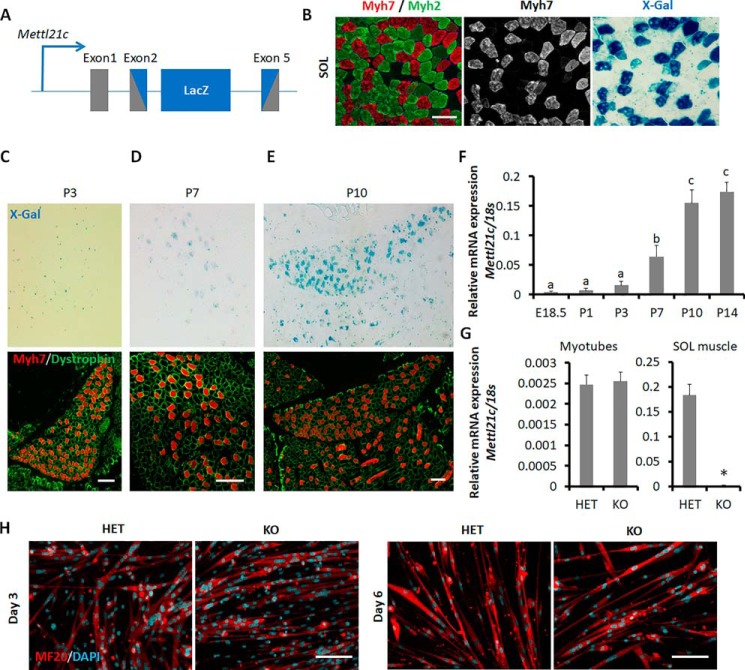
Figure 1.
Dynamic expression of Mettl21c during muscle development and myogenesis. A, genetic targeting of Mettl21c through insertion of LacZ into exons 2–5. The gray box indicates the exon of Mettl21c gene, and the blue box indicates LacZ gene. B, immunostaining of Myh7 (type I myofibers, red) and Myh2 (type IIa myofibers, green) and X-Gal staining of serial sections of SOL muscles isolated from Mettl21cLacZ/+ mice. Scale bar: 100 μm. C–E, immunostaining of Myh7 (red) and dystrophin (green), and X-Gal staining of serial sections of SOL muscles of P3 (C), P7 (D), and P10 (E) Mettl21cLacZ/+ mice. Scale bar: 100 μm. F, qPCR analysis of Mettl21c in different developmental stage. Error bars represent mean + S.D. of six mice with three technical repeats. Different letters indicate p < 0.05 (one-way ANOVA). G, qPCR analysis of Mettl21c in myocytes differentiated for 3 days or SOL muscles isolated from Mettl21cLacZ/+ (HET) or Mettl21cLacZ/LacZ (KO) mice. Error bars represent mean + S.D. of five independent biological experiments with three technical repeats. * indicates p < 0.05 (Student's t test). H, immunostaining of MF20 (myosin heavy chain, red) and DAPI (cyan) in differentiated myoblasts isolated from Mettl21cLacZ/+ mice at different days after differentiation induction. Scale bar: 50 μm.

