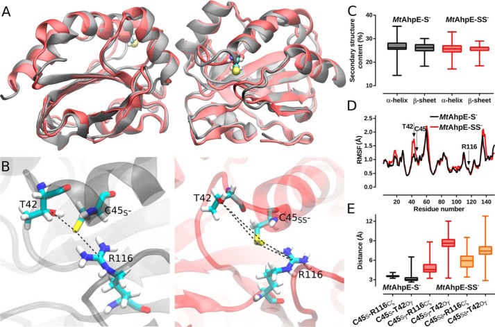
Figure 7.
Structural changes upon persulfidation in MtAhpE's active site. A, structural alignment of representative snapshots of the MtAhpE dimer obtained from MD simulations with Cys45 as thiolate (black) or as persulfide (red). B, comparison of thiolate and persulfide in Cys45 in the active site of MtAhpE depicting residues Thr42, Cys45 and Arg116. C, distribution of α-helix and β-sheet content (%) for both MD simulations. D, comparison of root mean square fluctuations (Å) in a per-residue basis calculated from thiolate in Cys45 (black) and persulfide in Cys45 (red) MD simulations. E, distribution of relevant active-site interactions. Selected distances are the same as highlighted in B.

