Figure 4.
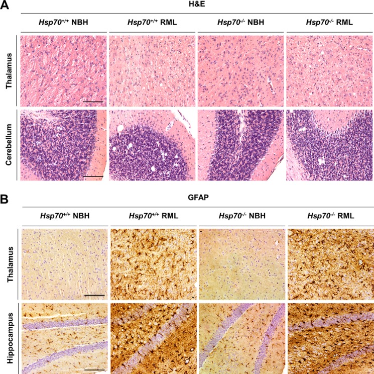
Histological changes in RML prion–infected Hsp70−/− and Hsp70+/+ mice. A, spongiform degeneration was analyzed in paraffin-embedded brain tissue slices from Hsp70−/− and Hsp70+/+ mice inoculated with RML prions. As controls, we used animals of both genotypes injected with NBH from a healthy animal. Slices were stained with H&E. B, gliosis was evaluated in fixed brain samples from Hsp70−/− and Hsp70+/+ mice inoculated with RML prions or NBH by probing with anti-glial fibrillary acidic protein (GFAP) antibody. Cell nuclei were simultaneously counterstained with hematoxylin. For the studies in this figure, we focused on the thalamus, hippocampus, and cerebellum, areas heavily affected by RML prion infection. Scale bars = 250 μm.

