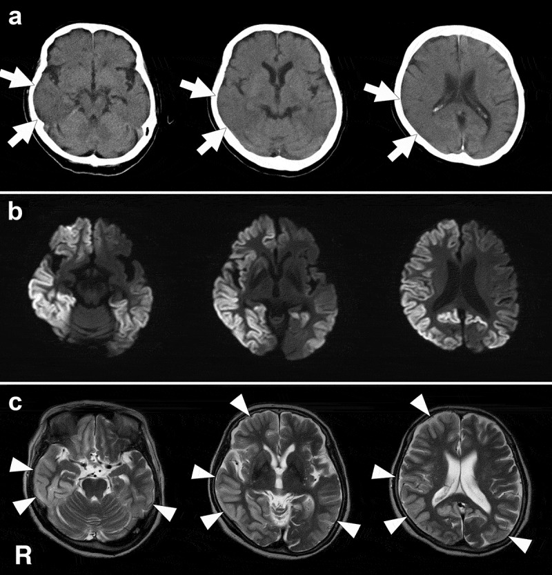
Figure 1.

Head computed tomography (CT) and magnetic resonance imaging (MRI) findings. These images were obtained 8 months after the disease onset. Clinically, facial mimicry and pathological laughing were remarkable at this stage. a: CT images show edematous and swelling findings in the right cerebral hemisphere, particularly in the temporal and parietal lobes (arrows). In the right temporal and parietal cortices, the sulcus shows narrowing and the corticomedullary junction is generally indistinct. b: Diffusion weighted MRI shows widespread gyriform hyperintensity in the right cerebral hemisphere and left parietal and occipital lobes, except in the medial occipital regions. c: T2-weighted images show hyperintense regions with swelling (arrowheads), and the regions correspond to those that were hyperintense on diffusion weighted MRI. R, right side.
