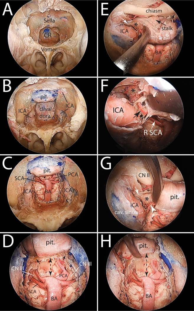
FIG. 2.

Stepwise cadaveric dissection demonstrating the EEA to the basilar apex. A: Expanded EEA with bilateral middle turbinectomies, posterior nasal septectomy, and a large sphenoidotomy performed to expose the sella and clival recess. B: Bone drilling of the ventral skull base has been completed from the planum sphenoidale superiorly to the floor of the sphenoid sinus inferiorly. C: Lateral extensions of bone drilling are the paraclival ICAs. The clival dura has been opened and the basilar artery is exposed, as is its terminal quadrifurcation. D: Close-up view of the basilar apex region showing the remaining dorsum sella bone (small black arrows,) and the relationship between the basilar apex, the oculomotor nerves, and the floor of the third ventricle. The dashed double-headed arrow shows the space available superior to the basilar apex without a PT. E: PT starts with opening the dura of the sellar region and developing the plane between the pituitary capsule medially and the medial dural wall of the cavernous sinus. This exposes the posterior clinoid process (asterisk). Small white arrow marks the attachment of posterior petroclinoid ligament to the posterior clinoid process. F: Magnified view of the image in E showing the inferior hypophyseal artery originating from the cavernous segment of the ICA (black arrow.) This small artery needs to be divided to allow for a full exposure of the posterior clinoid process (asterisk.) G: Medial mobilization of the pituitary to expose the inferior hypophyseal artery (white arrowhead,) the posterior clinoid process (asterisk), and the posterior petroclinoid ligament attaching to its posterolateral aspect (white arrow.) H: Bilateral transposition of the pituitary is completed with resection of the dorsum sellae and bilateral posterior clinoid processes. Note the increased space available superior to the basilar apex (dashed double-headed arrow) and compare with that shown in D. cav. = cavernous; CN = cranial nerve; CR = clival recess; pit. = pituitary; R = right.
