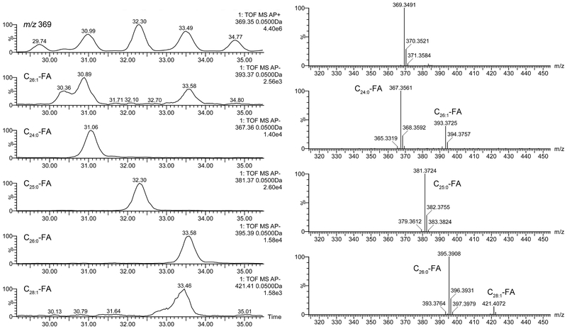
Figure 8. C18-UPLC/APCI-HRMS analysis of cholesteryl esters detected in tarsal plate extracts of a male mouse in positive and negative ion modes. The analysis is based on spontaneous in-source fragmentation of CE which produces an ion m/z 369.3521 (Chl − H2O + H)+ in PIM, and corresponding FA fragments (FA – H)− in NIM.
Left panels. A fragment of an extracted ion chromatograms of ions m/z 369.35 (PIM) and five FA fragments m/z 393.37, 367.36, 381.37, 395.39, and 421.41 (NIM).
Right panels. High resolution mass spectra of fragments of cholesterol (m/z 369.35; PIM) and three FA fragments of CE m/z 393.37, 367.36, 381.37, 395.39, and 421.41 (NIM).

