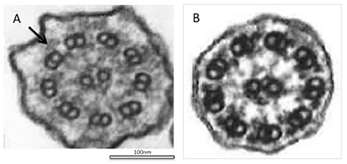
Figure 3.
Electron microscopy of cilia from bronchial mucosa. A: A transmission electron microscopic analysis shows a defect in the outer dynein arm in the cilia (arrow). B: Normal structure of cilia, characterized by a typical ’9+2’ structure with nine outer microtubule doublets and a central pair of microtubules.

