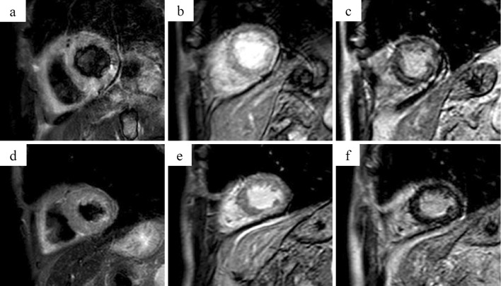
Figure 2.
Cardiac magnetic resonance imaging. Day 1, T2-weighted short-tau inversion recovery (STIR) black-blood (BB) (T2w-STIR-BB) MRI showed diffuse high signal intensity (SI) equal to or greater than the spleen SI. (a) Day 1, early gadolinium-enhanced (EGE) imaging showed diffuse hyper-enhancement of the myocardium. (b) Day 1, late gadolinium-enhanced (LGE) imaging showed diffuse patchy enhancement. (c) Given the above, myocarditis with diffuse myocardial edema was diagnosed. Day 11, T2w-STIR-BB (d) and EGE (e) images showed improvement of edematous findings, and late gadolinium enhancement (f) of the myocardium was decreased.

