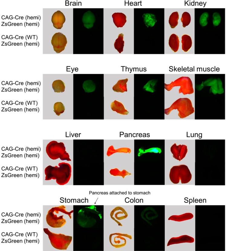
Figure 2.
Fluorescent images of individual tissues dissected from representative 3–4 week old rats of various genotypes. Images of fresh tissue were taken immediately after euthanasia at 1X magnification using a dissection microscope under bright field (images to left). Fluorescent images were taken at 1X using 488 nm excitation with an emission filter of 503 to 563 nm (images to right). Tissues from a double hemizygous rat are shown on top and tissues from a littermate that carries only the ZsGreen transgene and not the NCre transgene are shown on the bottom.

