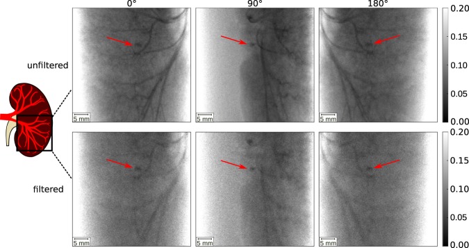
Figure 2.
MuCLS X-ray projection images filtered and unfiltered CTs at projection angles 0°, 90° and 180°. The kidney stone is indicated by a red arrow. In the unfiltered projection images, the iodine filled blood vessels are clearly visible together with the kidney stone. In the filtered projection images, the X-ray attenuation of the iodine contrast agent is reduced, yet still the differentiation of iodine and the kidney stone is difficult. The gray scales of the projection images show the relative transmission of the X-ray beam.

